Understanding Fibroids
What Are Fibroids?
Fibroids, also known as uterine leiomyomas, are non-cancerous growths that develop in the muscular wall of the uterus. They can vary significantly in size, shape, and number, ranging from small seedlings to large masses that can distort the shape of the uterus. While many women with fibroids experience no symptoms and may remain unaware of their presence, others may face a range of complications, including heavy menstrual bleeding, pelvic pain, and pressure symptoms that can impact daily life.
The exact cause of fibroids remains unclear, though research suggests that hormonal factors, particularly estrogen and progesterone, play a significant role in their development. These hormones stimulate the growth of the uterine lining during the menstrual cycle, and fibroids may respond similarly by enlarging in size. Genetic factors may also contribute, as fibroids often run in families. Additionally, certain environmental factors, such as diet and exposure to specific chemicals, are being explored for their potential influence on fibroid growth.
Fibroids can be classified into several types based on their location within the uterus. Submucosal fibroids grow just beneath the inner lining of the uterus and can lead to significant menstrual irregularities. Intramural fibroids are located within the muscular wall of the uterus and may cause pressure and discomfort. Subserosal fibroids develop on the outer surface of the uterus and can sometimes extend to nearby organs, leading to additional complications. Understanding the type and location of fibroids is crucial in determining the appropriate management strategies.
Women may experience a variety of symptoms associated with fibroids, although some may remain asymptomatic. Common symptoms include heavy menstrual bleeding, prolonged periods, pelvic pressure, and pain during intercourse. In some cases, fibroids can lead to complications such as anemia due to excessive bleeding or urinary issues if they press against the bladder. It is essential for women experiencing these symptoms to seek medical evaluation, as timely intervention can improve quality of life and prevent further complications.
In conclusion, while fibroids are common and often benign, they can significantly affect a woman’s health and wellbeing. Awareness of the nature of fibroids, their potential symptoms, and the importance of medical evaluation is vital for women seeking to understand their reproductive health. Proactive management strategies, including lifestyle modifications, medical treatments, and surgical options, can empower women to take control of their health and mitigate the impact of fibroids on their lives.
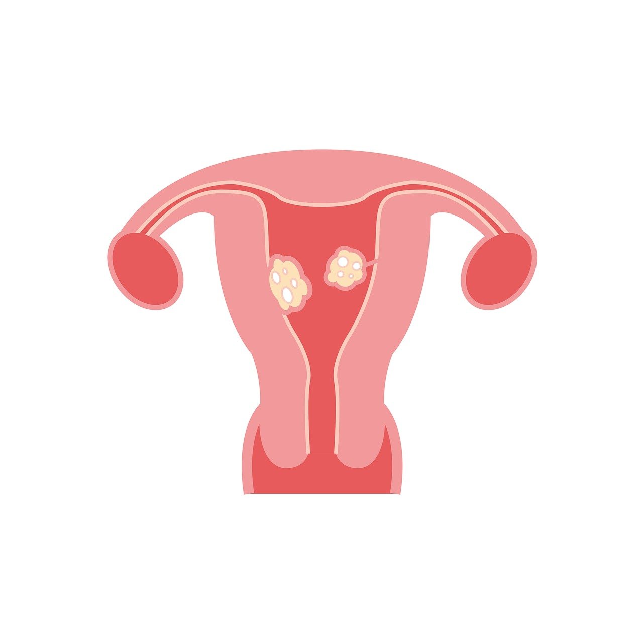
Types of Fibroids
Fibroids, also known as uterine leiomyomas, are non-cancerous growths that develop within the muscular wall of the uterus. They are classified into several types based on their location within the uterine structure. Understanding these types is crucial for devising effective management strategies. The main types include intramural, subserosal, submucosal, and pedunculated fibroids, each presenting unique characteristics and implications for women’s health.
Intramural fibroids are the most common type, occurring within the uterine wall itself. These fibroids can vary in size and may cause a range of symptoms, including heavy menstrual bleeding, pelvic pain, and pressure symptoms. Their location within the uterus can lead to significant distortion of the uterine cavity, potentially impacting fertility and complicating pregnancy. Women experiencing symptoms related to intramural fibroids may benefit from a range of treatment options, including medication, minimally invasive procedures, or surgical interventions.
Subserosal fibroids develop on the outer surface of the uterus and can grow quite large, often extending outward from the uterine wall. While many women with subserosal fibroids may remain asymptomatic, larger fibroids can exert pressure on adjacent organs, leading to discomfort and urinary or bowel issues. These fibroids may also be detected incidentally during imaging studies. Management of subserosal fibroids typically focuses on alleviating symptoms and may involve monitoring, medication, or surgical removal if they significantly affect quality of life.
Submucosal fibroids are located just beneath the endometrial lining of the uterus and can protrude into the uterine cavity. These fibroids are particularly concerning for women who are trying to conceive, as they can interfere with implantation and lead to complications during pregnancy. Symptoms may include heavy menstrual bleeding and reproductive issues. Treatment for submucosal fibroids often involves procedures aimed at preserving fertility, such as hysteroscopic myomectomy, which allows for the removal of the fibroid through the cervix.
Pedunculated fibroids are attached to the uterus by a stalk, or peduncle, and can be either subserosal or submucosal. These fibroids may cause unique symptoms depending on their location and can sometimes cause torsion, leading to acute pain. The management of pedunculated fibroids may include observation, medication, or surgical options depending on their size, symptoms, and impact on the individual’s health. Understanding the different types of fibroids empowers women to engage in informed discussions with their healthcare providers, facilitating tailored treatment plans that address their specific needs and concerns.
Causes and Risk Factors
Fibroids, also known as uterine leiomyomas, are non-cancerous growths that develop in the muscular wall of the uterus. While the exact cause of fibroids remains unclear, several factors have been identified that may contribute to their development. Hormonal influences, particularly estrogen and progesterone, play a significant role in the growth of fibroids. These hormones stimulate the uterine lining during the menstrual cycle, and fluctuations in their levels can lead to the proliferation of fibroid cells. This connection suggests that fibroids may be more prevalent during reproductive years when hormone levels are at their peak.
Genetics is another critical factor in the development of fibroids. Research indicates that women with a family history of fibroids are more likely to develop them themselves. Specific genetic mutations may predispose individuals to fibroid formation, highlighting the potential hereditary nature of this condition. Understanding the genetic components associated with fibroids can provide insights into risk assessment and aid in developing targeted treatments for affected individuals.
Environmental factors also contribute to the risk of developing fibroids. Obesity has been identified as a significant risk factor, as excess body fat can alter hormone levels and lead to increased estrogen production. Additionally, exposure to certain environmental toxins, such as endocrine disruptors found in plastics and personal care products, may also influence fibroid development. This underscores the importance of considering lifestyle and environmental factors when addressing the causes of fibroids.
Age and race are significant risk factors as well. Fibroids are most commonly diagnosed in women in their 30s and 40s, with the incidence decreasing after menopause when hormone levels decline. Furthermore, studies have shown that African American women are at a higher risk of developing fibroids compared to women of other racial and ethnic groups. This disparity suggests that both biological and socio-economic factors may contribute to the higher prevalence of fibroids in this population.
Understanding the various causes and risk factors associated with fibroids is essential for effective management and prevention strategies. By identifying individuals at higher risk, healthcare professionals can provide tailored recommendations aimed at reducing the likelihood of fibroid development. Moreover, increased awareness of these factors can empower individuals to make informed lifestyle choices that may mitigate their risk, ultimately leading to improved health and wellness in the face of this common condition.
Symptoms of Fibroids
Fibroids, also known as uterine leiomyomas, are noncancerous growths that develop in the muscular wall of the uterus. They vary significantly in size, shape, and location, which can lead to a diverse range of symptoms. Understanding these symptoms is crucial for early detection and management. While some women with fibroids may remain asymptomatic, others may experience a variety of issues that can significantly impact their quality of life.
One of the most common symptoms associated with fibroids is heavy menstrual bleeding, known medically as menorrhagia. Women may notice an increase in the volume and duration of their periods, sometimes accompanied by the passing of large blood clots. This condition can lead to anemia and fatigue, as the body struggles to replace lost blood. The severity of bleeding can vary, and in some cases, it may necessitate medical intervention or surgical options to alleviate the symptoms.
In addition to heavy bleeding, many women report experiencing pelvic pain or pressure. This discomfort can result from the fibroids pressing against surrounding organs, such as the bladder or rectum. The sensation may manifest as a constant ache or a sharp pain, particularly during menstrual cycles or physical activity. It is important for women to differentiate between normal menstrual discomfort and pain that may indicate a complication, such as torsion or degeneration of the fibroids.
Another symptom that warrants attention is the increased frequency of urination. When fibroids grow in size, they can exert pressure on the bladder, leading to a heightened need to urinate or a feeling of incomplete bladder emptying. This can disrupt daily activities and sleep patterns, causing further stress and discomfort. Additionally, some women may experience difficulties with bowel movements, including constipation or discomfort, due to the pressure from fibroids on the rectal area.
Lastly, some women with fibroids may encounter complications related to fertility. While fibroids alone do not always impede conception, they can interfere with implantation or lead to complications during pregnancy. Symptoms related to fertility can be subtle, but they may include irregular periods or difficulty conceiving. Women experiencing these issues should consult healthcare providers to explore potential connections to fibroids and discuss appropriate treatment options.
Recognizing the symptoms of fibroids is essential for timely diagnosis and effective management. Women experiencing any combination of heavy menstrual bleeding, pelvic pain, urinary frequency, or fertility challenges should seek medical advice. Early intervention can lead to better outcomes and improved quality of life, empowering women to take control of their health and well-being in the face of fibroid-related challenges.
Diagnosis and Evaluation
Medical History and Physical Examination
Medical history and physical examination play crucial roles in the diagnosis and management of fibroids. A comprehensive medical history involves gathering detailed information about a patient’s symptoms, menstrual cycle, and any prior treatments. Patients are encouraged to describe the onset, duration, and severity of their symptoms, such as heavy menstrual bleeding, pelvic pain, or pressure. Additionally, understanding family history is essential, as fibroids can have a hereditary component. A thorough medical history not only aids in the accurate diagnosis but also helps in tailoring treatment options that align with the patient’s specific needs.
During the physical examination, healthcare providers perform a pelvic exam to assess the size and location of fibroids. This examination allows clinicians to identify any abnormalities in the uterus and surrounding tissues. In some cases, the healthcare provider may also recommend imaging studies, such as ultrasound or MRI, to obtain a clearer picture of the fibroids. These imaging techniques provide valuable information regarding the number, size, and configuration of the fibroids, guiding the development of an effective management plan.
Understanding the impact of fibroids on a patient’s quality of life is also an integral part of the assessment process. Patients often experience a range of emotional and psychological effects due to their symptoms, which can lead to feelings of anxiety or depression. The healthcare provider should take the time to discuss these aspects with the patient, fostering an environment where concerns can be openly addressed. This approach not only aids in building a strong patient-provider relationship but also ensures that all factors influencing the patient’s health and well-being are considered.
In some cases, additional diagnostic procedures may be warranted if the initial examination indicates complications or if the diagnosis remains uncertain. Procedures such as hysteroscopy or endometrial biopsy can be performed to gather further information. These minimally invasive techniques allow for direct visualization of the uterine cavity and can help rule out other potential causes of symptoms, ensuring a more accurate diagnosis. It is essential that patients understand the purpose of these tests and feel empowered to participate in the decision-making process regarding their care.
Ultimately, a thorough medical history and physical examination form the foundation of effective fibroid management. By engaging in open communication and providing comprehensive evaluations, healthcare providers can develop personalized treatment strategies that address the unique challenges faced by each patient. This collaborative approach not only improves clinical outcomes but also enhances the overall quality of life for individuals dealing with fibroids, paving the way toward wellness and relief.
Imaging Techniques
Imaging techniques play a crucial role in the diagnosis and management of fibroids, providing essential information about their size, location, and impact on surrounding structures. These non-invasive and minimally invasive imaging modalities enable healthcare providers to tailor treatment strategies effectively. Among the most common techniques used are ultrasound, magnetic resonance imaging (MRI), and computed tomography (CT). Each method has unique strengths, allowing practitioners to select the most appropriate one based on individual patient circumstances.
Ultrasound is often the first-line imaging technique for evaluating fibroids due to its accessibility, cost-effectiveness, and ability to visualize the uterus in real time. Transabdominal and transvaginal ultrasound can both be employed to assess fibroids’ characteristics, including their number and size. The transvaginal approach is particularly useful for detailed imaging of the uterus and can provide better visualization of smaller fibroids. Additionally, Doppler ultrasound can assess blood flow to the fibroids, offering insights into their potential for causing symptoms.
Magnetic resonance imaging (MRI) provides a more detailed view of fibroids and the surrounding pelvic structures compared to ultrasound. It is particularly valuable in complex cases where the relationship of fibroids to other organs needs to be assessed, or when the diagnosis is uncertain. MRI can differentiate between types of fibroids and other pelvic masses, as well as provide information regarding the extent of degeneration within the fibroids. This imaging technique is beneficial for planning surgical interventions, as it allows for precise mapping of fibroid locations and sizes.
Computed tomography (CT) is less commonly used for fibroid assessment due to its exposure to ionizing radiation and limited soft tissue contrast compared to MRI. However, it can be useful in specific scenarios, such as when evaluating complications related to fibroids, including potential obstruction of urinary structures or identification of associated masses. CT can also assist in surgical planning by providing a comprehensive view of the pelvic anatomy, although MRI remains the preferred choice in most cases.
In summary, imaging techniques are integral to the effective management of fibroids, guiding diagnosis and influencing treatment decisions. Healthcare providers must consider the advantages and limitations of each modality to select the most suitable option for their patients. By leveraging these advanced imaging technologies, clinicians can enhance their understanding of fibroid characteristics and develop personalized treatment plans that address the unique needs of each individual.
Differential Diagnosis
Differential diagnosis is a crucial aspect of managing fibroids, as it allows healthcare providers to distinguish between fibroids and other conditions that may present with similar symptoms. The presence of uterine fibroids often leads to a variety of symptoms, including heavy menstrual bleeding, pelvic pain, and pressure symptoms. However, these symptoms can also be indicative of other uterine abnormalities such as adenomyosis, endometriosis, or even pelvic inflammatory disease. A comprehensive evaluation is essential to ensure proper diagnosis and treatment.
A thorough patient history is the first step in the differential diagnosis process. Clinicians should inquire about the severity and duration of symptoms, menstrual cycle regularity, and any associated factors such as pain during intercourse or changes in urinary habits. Additionally, a detailed medical history that includes any previous gynecological conditions, surgeries, or family history of reproductive health issues can provide valuable context. This information assists healthcare providers in formulating a differential diagnosis that considers the most likely conditions based on the patient’s profile.
Physical examination, particularly a pelvic exam, plays a significant role in the diagnostic process. During the pelvic exam, the healthcare provider can assess for any abnormalities in the size and shape of the uterus, as well as any palpable masses. Ultrasound imaging is often the first-line imaging modality utilized to visualize fibroids and assess their size and location. However, in certain cases, advanced imaging techniques such as MRI may be warranted to provide a clearer picture of the uterine anatomy and to rule out other potential diagnoses, such as uterine cancer or complex ovarian cysts.
In addition to imaging, laboratory tests may also be employed to aid in the differential diagnosis. Blood tests to check for anemia can be particularly useful, especially in cases where heavy menstrual bleeding is reported. Hormonal assessments may also help in determining if hormonal imbalances are contributing to the symptoms. By combining clinical findings, imaging results, and laboratory data, healthcare providers can arrive at a more accurate diagnosis, guiding them toward the most appropriate treatment options.
Finally, it is essential for patients to understand the importance of clear communication with their healthcare providers throughout the diagnostic process. Engaging in discussions about symptoms and concerns can lead to a more accurate diagnosis and a tailored treatment plan. This collaborative approach not only empowers patients but also enhances the overall quality of care. By recognizing the nuances of differential diagnosis, individuals affected by fibroids can take proactive steps toward effective management and improved well-being.
When to Seek Medical Advice
Recognizing the appropriate time to seek medical advice is crucial for individuals dealing with fibroids. While many people may experience benign symptoms, there are specific indicators that warrant professional evaluation. If you encounter persistent pelvic pain or discomfort that interferes with daily activities, it is essential to consult a healthcare provider. These symptoms could signify a more significant issue requiring intervention. Additionally, if menstrual bleeding becomes excessively heavy or prolonged, leading to anemia or fatigue, it is vital to seek medical advice promptly.
Changes in urinary or bowel habits can also be a sign that medical attention is necessary. Increased frequency of urination, difficulty emptying the bladder, or constipation may indicate that fibroids are pressing on nearby organs. These symptoms can significantly affect quality of life and may necessitate further investigation. A healthcare professional can assess the situation and recommend appropriate imaging or tests to determine the underlying cause of these changes.
Another critical factor to consider is the presence of abdominal swelling or a noticeable bulge in the lower abdomen. While some degree of abdominal discomfort can be typical with fibroids, sudden changes in size or shape may indicate complications that require medical evaluation. This may include torsion or degeneration of the fibroid, which can lead to severe pain and other complications. In such cases, timely medical intervention is critical to avoid further health issues.
Individuals who are trying to conceive should also be vigilant about their symptoms and seek medical advice if they suspect fibroids may be impacting their fertility. Fibroids can potentially interfere with implantation or increase the risk of miscarriage, and early intervention can be beneficial. A thorough evaluation by a specialist can provide insight into the impact of fibroids on reproductive health and outline potential treatment options that may enhance fertility.
Lastly, it is essential to establish open communication with healthcare providers about any concerns related to fibroids. Regular check-ups and discussions about symptoms can facilitate early detection of changes in condition. Empowering oneself with knowledge about fibroids and understanding when to seek medical advice can significantly influence the management of the condition, ultimately leading to improved health outcomes and enhanced quality of life.
No responses yet