Introduction to Pedunculated Fibroids
Definition of Pedunculated Fibroids
Pedunculated fibroids are a specific type of uterine fibroid characterized by their unique attachment to the uterus via a stalk or peduncle. This stalk allows the fibroid to hang away from the uterine wall, differentiating it from other types of fibroids that are embedded directly within the uterine tissue. Pedunculated fibroids can be found either on the outer surface of the uterus, known as subserosal fibroids, or within the uterine cavity, referred to as submucosal fibroids. Their location plays a significant role in the symptoms they may cause and the treatment options available.
The formation of pedunculated fibroids is attributed to the growth of smooth muscle cells and connective tissue in the uterus. These fibroids are composed of muscle and fibrous tissue and can vary in size, ranging from small nodules to large masses. While the exact cause of fibroid development remains unclear, factors such as hormonal influences, particularly estrogen, and genetic predispositions are believed to play a significant role. Understanding the biological underpinnings of pedunculated fibroids is essential for both clinicians and patients in managing their implications.
Symptoms associated with pedunculated fibroids can vary widely depending on their size, location, and whether they are causing any obstruction or pressure on surrounding organs. Some individuals may experience heavy menstrual bleeding, pelvic pain, or pressure symptoms, while others may remain asymptomatic. The positional nature of pedunculated fibroids can lead to unique challenges, such as torsion of the peduncle, which can result in acute pain and require urgent medical intervention.
Diagnosis of pedunculated fibroids typically involves imaging techniques such as ultrasound or magnetic resonance imaging (MRI). These modalities allow for a clear visualization of the fibroids, helping to determine their size, location, and potential impact on reproductive health. Accurate diagnosis is crucial, as it informs the treatment approach, which can range from observation to surgical intervention, depending on the severity of symptoms and the individual’s reproductive goals.
Management of pedunculated fibroids requires a tailored approach that considers the patient’s unique circumstances. Treatment options may include medication to manage symptoms, minimally invasive procedures, or surgical options, such as myomectomy or hysterectomy, depending on the severity and impact of the fibroids. Education and support are vital for individuals affected by pedunculated fibroids, as understanding their condition empowers patients to make informed decisions regarding their health and treatment options.
Types of Fibroids
Fibroids, also known as uterine leiomyomas, are benign tumors that develop in the muscle tissue of the uterus. These growths can vary significantly in size, shape, and location, leading to a variety of classifications. Among the different types of fibroids, pedunculated fibroids are distinct due to their unique attachment to the uterine wall via a stalk or peduncle. This feature not only influences their growth patterns but also impacts the symptoms and potential complications they may cause.
Pedunculated fibroids can be further categorized into two main types: pedunculated subserosal fibroids and pedunculated intramural fibroids. Pedunculated subserosal fibroids arise from the outer surface of the uterus and extend outward, often leading to a noticeable bulge on the abdomen. These fibroids are typically more likely to be symptomatic, as their growth can exert pressure on surrounding organs, leading to discomfort or pain. Conversely, pedunculated intramural fibroids develop within the uterine wall itself and can also project outward. While they share a similar stalk with subserosal fibroids, their location can lead to different clinical manifestations.
The size of pedunculated fibroids can greatly influence the symptoms experienced by individuals. Smaller fibroids may remain asymptomatic, while larger fibroids often lead to significant issues such as pelvic pain, heavy menstrual bleeding, or complications during pregnancy. The presence of a peduncle can also alter the dynamics of blood flow to the fibroid, potentially resulting in degeneration or torsion. Torsion occurs when the fibroid twists around its stalk, leading to acute pain and requiring immediate medical attention. Understanding the implications of size and type is crucial for effective management.
Diagnosis of pedunculated fibroids typically involves imaging studies such as ultrasound or MRI, which can provide detailed information about their size, location, and type. These diagnostic methods are essential for differentiating between various fibroid types and other potential conditions, such as ovarian cysts or malignancies. Once accurately diagnosed, treatment options can be tailored to the individual’s needs, considering factors such as symptom severity, size of the fibroid, and reproductive plans.
Management strategies for pedunculated fibroids range from conservative monitoring to surgical interventions. In cases where symptoms are mild or absent, a watchful waiting approach may be appropriate. However, for those experiencing significant discomfort or complications, surgical options, including myomectomy or hysterectomy, may be considered. The choice of treatment depends on various factors, including the patient’s age, desire for future fertility, and overall health. By understanding the types of fibroids and their implications, individuals can make informed decisions regarding their care and treatment options.
Importance of Understanding Pedunculated Fibroids
Understanding pedunculated fibroids is crucial for anyone affected by them, as these benign tumors can significantly impact health and quality of life. Pedunculated fibroids are unique in their structure, characterized by a stalk-like attachment to the uterus, which can lead to various symptoms and complications. This distinction is vital for proper diagnosis and treatment. Awareness of these fibroids allows individuals to make informed decisions regarding their health and seek appropriate medical care.
One of the primary reasons for understanding pedunculated fibroids is the potential for symptom development. While some individuals may remain asymptomatic, others may experience pain, abnormal bleeding, or pressure symptoms depending on the fibroid’s size and location. Recognizing these symptoms early can lead to timely medical intervention, preventing further complications such as torsion of the fibroid or anemia due to excessive bleeding. Understanding the nature of pedunculated fibroids empowers individuals to communicate effectively with healthcare providers about their experiences and concerns.
Additionally, understanding pedunculated fibroids can influence treatment options. Healthcare providers often recommend a range of management strategies, from watchful waiting to surgical interventions, based on the fibroid’s characteristics and the patient’s symptoms. Being knowledgeable about the existence and implications of pedunculated fibroids allows individuals to engage in discussions about their treatment plans actively. This engagement fosters a collaborative relationship with healthcare professionals, ensuring that patients receive personalized care tailored to their specific circumstances.
Furthermore, awareness of pedunculated fibroids plays a significant role in addressing misconceptions and reducing stigma associated with fibroid-related conditions. Many individuals may feel embarrassed or isolated due to their symptoms, leading to reluctance in seeking help. By promoting understanding and education about pedunculated fibroids, we can create a supportive environment where individuals feel empowered to discuss their health openly. This not only aids in personal health management but also contributes to broader societal awareness and advocacy for women’s health issues.
In conclusion, the importance of understanding pedunculated fibroids cannot be overstated. They represent a significant aspect of gynecological health that affects many individuals. By recognizing the symptoms, exploring treatment options, and fostering open discussions, individuals can take charge of their health. This knowledge equips them to navigate their experiences with pedunculated fibroids, ultimately enhancing their quality of life and promoting overall well-being.
Anatomy and Physiology
Uterine Anatomy
The uterus, a key organ in the female reproductive system, is situated in the pelvic cavity and plays an essential role in menstruation, pregnancy, and childbirth. Its anatomy is complex, comprising three main layers: the endometrium, myometrium, and perimetrium. The endometrium, the innermost layer, is responsible for the cyclical changes that occur during the menstrual cycle, providing a suitable environment for embryo implantation. The myometrium, a thick layer of smooth muscle, facilitates contractions during labor and helps expel menstrual fluid. The perimetrium, the outermost layer, serves as a protective covering for the uterus. Understanding these layers is crucial for comprehending how conditions such as pedunculated fibroids can develop and affect uterine function.
Pedunculated fibroids are non-cancerous growths that can emerge from the uterine wall or from the endometrial lining. Their unique structure distinguishes them from other types of fibroids, as they are attached to the uterus by a stalk or peduncle. This stalk can vary in length, and the fibroid can reside within the uterine cavity or extend outward. The location of pedunculated fibroids can significantly influence the symptoms experienced by the individual, as they may cause pressure on adjacent organs or disrupt normal menstrual function. The growth of these fibroids is often linked to hormonal imbalances, particularly estrogen, which plays a vital role in the growth and regulation of uterine tissue.
The vascular supply to the uterus is critical for maintaining its health and function. The uterine arteries, which branch from the internal iliac arteries, provide oxygenated blood to the uterus, while the ovarian arteries supply blood to the ovaries. This rich blood supply not only supports normal uterine processes but also facilitates the growth of fibroids. As pedunculated fibroids develop, they may receive blood flow through both the uterine and ovarian arteries, which can contribute to their growth and the symptoms they produce. Understanding the vascular anatomy of the uterus is essential when evaluating the potential impact of fibroids on overall reproductive health.
The position of the uterus within the pelvic cavity also plays a significant role in the presentation of pedunculated fibroids. The uterus typically tilts forward (anteverted) or backward (retroverted), and its position can affect the symptoms experienced by individuals with fibroids. For example, a retroverted uterus may allow fibroids to exert more pressure on the bladder, leading to urinary symptoms. Conversely, a forward-tilting uterus may result in pressure on the rectum or surrounding structures. Recognizing how the anatomical position of the uterus interacts with the presence of fibroids is vital for accurate diagnosis and effective treatment planning.
In summary, a thorough understanding of uterine anatomy is essential for appreciating the implications of pedunculated fibroids on women’s health. The interplay between the uterine layers, vascular supply, and anatomical positioning provides insight into the development and symptomatology of these growths. This knowledge not only aids healthcare professionals in diagnosing and managing fibroids but also empowers patients to engage in informed discussions about their reproductive health. As the exploration of uterine anatomy continues, it remains a foundational aspect of understanding the complexities surrounding pedunculated fibroids.
How Fibroids Develop
Fibroids, also known as leiomyomas, are non-cancerous growths that develop in the muscular wall of the uterus. The formation of these fibroids is a complex process influenced by a combination of genetic, hormonal, and environmental factors. Understanding how fibroids develop is crucial for the effective management and treatment of this condition. Pedunculated fibroids, characterized by a stalk-like attachment to the uterus, arise from the same underlying mechanisms that govern the growth of other types of fibroids.
The development of fibroids typically begins with the proliferation of smooth muscle cells in the uterine wall. Certain genetic mutations have been identified that may predispose some women to develop fibroids. These mutations can lead to abnormal cell growth and division, creating a localized mass of muscle tissue. Additionally, the presence of estrogen and progesterone plays a significant role in this process, as these hormones stimulate the growth of the uterine lining and are known to promote fibroid development.
As fibroids grow, they may develop a pedunculated structure, which occurs when the fibroid forms on a stalk or stem. This stalk allows the fibroid to hang away from the uterus, potentially causing different symptoms compared to other types of fibroids. The size and location of pedunculated fibroids can vary widely, influencing how they affect the surrounding tissues and organs. Some women may experience discomfort or pain, while others may remain asymptomatic.
Environmental factors, including diet and lifestyle choices, can also contribute to the development and growth of fibroids. Research suggests that obesity, a diet high in red meat and low in fruits and vegetables, and exposure to certain chemicals may increase the risk of developing fibroids. These factors can interact with hormonal pathways, further enhancing the growth potential of fibroids. Understanding these environmental influences can aid in identifying at-risk individuals and guiding preventative strategies.
Finally, the interplay between genetic predisposition, hormonal influences, and environmental factors culminates in the formation of pedunculated fibroids. While the exact mechanisms remain an area of ongoing research, recognizing how fibroids develop provides valuable insights into their prevention and treatment. This knowledge empowers individuals to make informed decisions regarding their health, encourages early intervention, and fosters a better understanding of the broader implications of fibroid development on reproductive health.
Characteristics of Pedunculated Fibroids
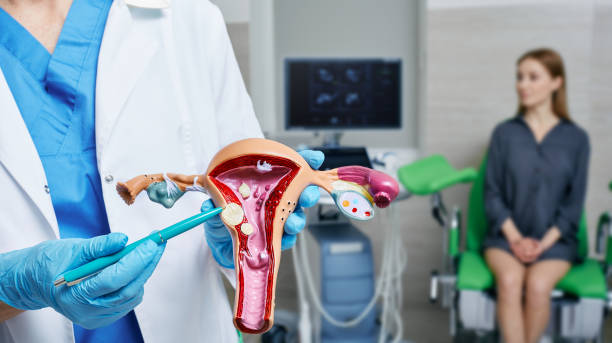
Pedunculated fibroids are a specific type of uterine fibroid characterized by their unique attachment to the uterine wall via a stalk or peduncle. This stalk can vary in length and can be located either within the uterine cavity or outside on the surface of the uterus. The distinctive structure of pedunculated fibroids is significant as it influences their behavior, symptoms, and potential complications. Understanding these characteristics is crucial for both patients and healthcare providers in making informed decisions regarding diagnosis and treatment.
One of the notable features of pedunculated fibroids is their mobility. Unlike other fibroid types that are firmly embedded in the uterine wall, pedunculated fibroids can sometimes move freely within the abdominal cavity. This movement can lead to a variety of symptoms, including abdominal pain or pressure, depending on the fibroid’s size and position. Additionally, because they can twist around their stalk, they may cause acute pain or complications such as torsion, which requires immediate medical attention.
Pedunculated fibroids can exhibit a range of sizes, from small nodules to larger masses that can significantly impact the surrounding organs. Their growth patterns may be influenced by hormonal factors, particularly estrogen, which can stimulate fibroid development. As a result, the presence of pedunculated fibroids can lead to symptoms such as heavy menstrual bleeding, pelvic discomfort, and urinary frequency, which may be exacerbated by their size and location. Patients should be aware of these potential symptoms, as they can significantly affect quality of life.
The imaging characteristics of pedunculated fibroids also play a crucial role in diagnosis. On ultrasound, these fibroids typically appear as well-defined masses with a clear stalk connecting them to the uterus. Magnetic resonance imaging (MRI) can provide more detailed information about the fibroid’s size, location, and relationship to surrounding structures. Accurate imaging is essential for distinguishing pedunculated fibroids from other types, such as subserosal or intramural fibroids, which may require different management strategies.
Treatment options for pedunculated fibroids vary depending on the severity of symptoms and the patient’s reproductive goals. While many pedunculated fibroids may not require intervention, those causing significant discomfort or complications may necessitate surgical removal. Options include laparoscopic surgery for smaller fibroids or more extensive procedures for larger masses. Understanding the characteristics of pedunculated fibroids aids in determining the most appropriate treatment approach, ensuring that patients receive personalized care tailored to their specific needs.
Causes and Risk Factors
Genetic Factors
Genetic factors play a crucial role in the development of pedunculated fibroids, which are benign tumors that arise from the smooth muscle tissue of the uterus. Research has indicated that hereditary predisposition is significant, with certain genetic mutations and familial patterns influencing the likelihood of fibroid formation. In particular, women with a family history of fibroids are more likely to develop these growths, suggesting that genetics can contribute to the overall risk profile of individuals. Understanding these genetic influences is essential for both patients and healthcare providers as they navigate the complexities of fibroid management.
Several key genes have been implicated in the pathogenesis of fibroids. Mutations in genes such as MED12, which is involved in the regulation of gene expression, have been frequently associated with fibroid development. Additionally, abnormalities in other genes that govern cell growth and division can lead to the uncontrolled proliferation of smooth muscle cells, contributing to the formation of fibroids. This evidence underscores the importance of genetic research in identifying not only the presence of fibroids but also their potential behavior and response to treatment.
The expression of various growth factors and hormones is also influenced by genetic predispositions, impacting the environment in which fibroids develop. For instance, the interplay between estrogen and progesterone, hormones that regulate the menstrual cycle, is critical in fibroid growth. Genetic variations can lead to differences in hormone receptor sensitivity, which may result in an increased growth response to these hormones. Understanding these hormonal interactions at a genetic level allows for a more nuanced approach to treatment, potentially leading to personalized therapies based on an individual’s genetic makeup.
Epigenetic factors, which involve changes in gene expression without altering the underlying DNA sequence, also contribute to the development of pedunculated fibroids. Environmental factors such as diet, stress, and exposure to certain chemicals can cause epigenetic modifications that may influence fibroid growth. This aspect of genetic research emphasizes the interplay between inherited traits and environmental influences, highlighting that while genetics play a significant role, lifestyle and external factors also contribute to the risk of developing fibroids.
In conclusion, genetic factors are instrumental in understanding the formation and progression of pedunculated fibroids. The interplay of hereditary predispositions, genetic mutations, hormonal influences, and epigenetic modifications creates a complex framework that shapes an individual’s risk profile. Continued research in this area is essential for developing targeted therapies and improving patient outcomes. As knowledge of the genetic underpinnings of fibroids expands, it will pave the way for more effective prevention strategies and personalized treatment options, ultimately enhancing the quality of life for those affected by these common uterine tumors.
Hormonal Influences
Hormones play a crucial role in the development and growth of pedunculated fibroids, which are benign tumors that form on the uterus. These fibroids, also known as uterine leiomyomas, are influenced significantly by sex hormones, particularly estrogen and progesterone. Estrogen is known to stimulate the growth of uterine tissue, and during a woman’s reproductive years, the fluctuating levels of this hormone can contribute to the enlargement of fibroids. Additionally, progesterone, which is produced during the second half of the menstrual cycle, can also affect fibroid growth, although its role is more complex and may vary depending on the individual.
The mechanism through which these hormones influence fibroid development involves their interaction with specific receptors present in the uterine tissue. Estrogen and progesterone bind to these receptors, triggering a cascade of biological responses that promote cellular proliferation and increase blood flow to the fibroid. As a result, fibroids can grow more rapidly during periods of hormonal surges, such as pregnancy, when estrogen levels are elevated. Understanding these hormonal dynamics is essential for grasping how fibroids may change in size and symptoms throughout a woman’s life.
Moreover, the hormonal environment is not static; it varies with different life stages and conditions. For instance, during menopause, the decline in estrogen levels typically leads to a reduction in fibroid size. This change underscores the importance of hormonal balance in managing fibroids and highlights why women experiencing hormonal fluctuations, such as those undergoing fertility treatments or hormone replacement therapy, may notice changes in their fibroid symptoms. The interplay of hormones can not only influence the size of fibroids but also affect the symptoms associated with them, such as pain and heavy menstrual bleeding.
In addition to endogenous hormones, external factors such as diet, lifestyle, and environmental exposures can also impact hormonal levels and, consequently, fibroid development. For example, obesity is linked to higher estrogen levels due to increased adipose tissue, which can produce estrogen. Similarly, certain dietary choices and exposure to endocrine disruptors found in some plastics and personal care products may contribute to hormonal imbalances. Recognizing these external influences is vital for women seeking to manage fibroid symptoms and overall health effectively.
In conclusion, understanding the hormonal influences on pedunculated fibroids is essential for both patients and healthcare providers. A thorough awareness of how estrogen and progesterone interact with fibroid growth can inform treatment options and lifestyle choices. Women experiencing fibroids should consider regular monitoring and discuss hormonal impacts with their healthcare providers, as this knowledge can lead to more personalized and effective management strategies for their condition.
Lifestyle and Environmental Factors
Lifestyle and environmental factors play a significant role in the development and management of pedunculated fibroids. These benign tumors, which arise from the muscle tissue of the uterus and are attached by a stalk, can be influenced by various aspects of a person’s daily life. Understanding these factors is essential for individuals seeking to minimize their risk or manage existing fibroids effectively.
Diet is one of the most critical lifestyle components that can affect fibroid growth. Research suggests that a diet high in red meat and low in fruits and vegetables may be associated with an increased risk of developing fibroids. Conversely, a diet rich in whole grains, dairy, and green leafy vegetables may help in reducing the risk. Additionally, maintaining a healthy weight is crucial, as obesity has been linked to higher estrogen levels, which can stimulate fibroid growth. Therefore, adopting a balanced diet can be an effective strategy for those concerned about pedunculated fibroids.
Physical activity is another vital lifestyle factor that can influence fibroid development. Regular exercise has been shown to lower estrogen levels and improve overall hormonal balance, which may contribute to a reduced risk of fibroid formation. Engaging in consistent physical activity can also help manage stress, which is known to exacerbate hormonal imbalances. Incorporating aerobic exercises, strength training, and flexibility workouts into one’s routine can provide both physical and mental health benefits, making it a crucial aspect of fibroid management.
Environmental factors, including exposure to endocrine-disrupting chemicals, can also impact the growth and development of pedunculated fibroids. Chemicals found in certain plastics, personal care products, and pesticides can mimic estrogen and disrupt hormonal balance. Individuals who are concerned about fibroids should be mindful of their exposure to these substances by choosing organic products when possible, using glass or stainless steel containers, and selecting personal care items that are free from harmful chemicals. By minimizing exposure to these environmental toxins, individuals may help reduce their risk of developing fibroids.
Stress management is equally important in the context of fibroid health. Chronic stress can lead to hormonal imbalances, which may contribute to the growth of fibroids. Techniques such as mindfulness, meditation, yoga, and deep-breathing exercises can be effective in managing stress levels. Additionally, fostering social connections and seeking support from friends, family, or support groups can enhance emotional well-being. By addressing lifestyle and environmental factors, individuals can take proactive steps toward understanding and managing pedunculated fibroids, ultimately improving their overall health and quality of life.

2 Responses
Thank you very much for the information. If I may ask can one fully recover from pedunculated fibroid?
Yes, it is possible to fully recover from pedunculated fibroids, especially with timely and appropriate treatment.
Treatment options:
Treatment depends on factors like size, location, symptoms, and overall health. Common options include:
1. Watchful waiting (monitoring)
2. Hormonal therapies
3. Uterine artery embolization (UAE)
4. Myomectomy (surgical removal)
5. Hysterectomy (removal of the uterus)
6. Laparoscopic or robotic surgery
7. High-intensity focused ultrasound (HIFU)
Recovery prospects:
With proper treatment, many women experience significant improvement or complete resolution of symptoms. Factors influencing recovery include:
1. Size and location of the fibroid
2. Severity of symptoms
3. Treatment effectiveness
4. Overall health
Post-treatment outcomes:
After treatment, women may experience:
1. Reduced or eliminated symptoms (pain, bleeding, pressure)
2. Improved quality of life
3. Enhanced fertility (if fibroids were affecting reproductive health)
4. Reduced risk of complications (e.g., anemia, infection)
Natural remedies and complementary therapies:
Some women explore alternative approaches, such as:
1. Dietary changes
2. Herbal supplements (e.g., turmeric, ginger)
3. Acupuncture
4. Yoga and stress management
While these may help alleviate symptoms, consult your healthcare provider before using them as primary treatment.
Consult a healthcare professional:
For personalized guidance and optimal outcomes, consult a gynecologist or healthcare provider. They will assess your specific situation and recommend the best course of treatment.